Back to blog list 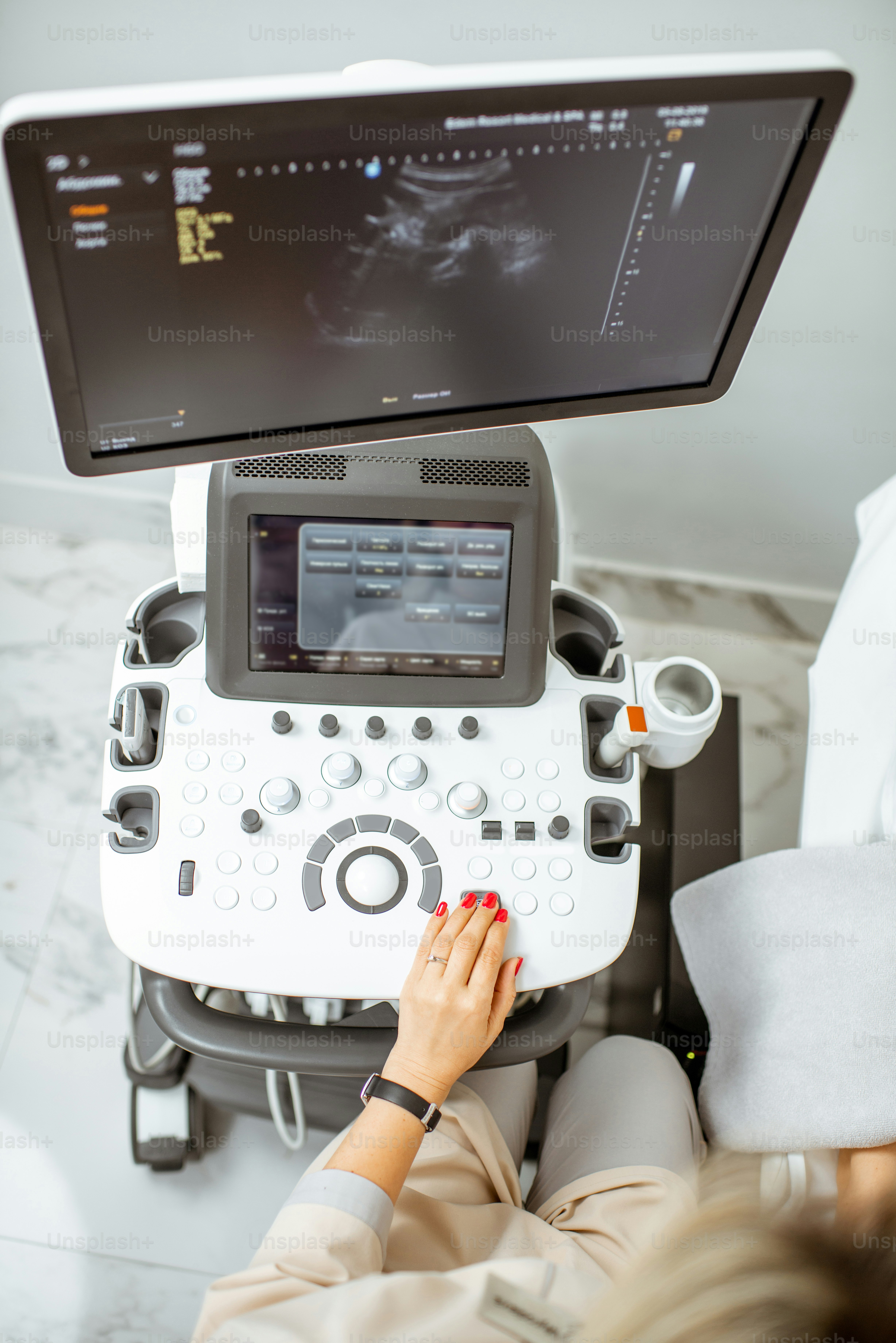
Ultrasound Scans: The Difference Between 2D, 3D, 4D, and 5D
• Tim Kelas Antenatal & Postnatal
Ultrasound diagnostics now span from classic 2D to spectacular 5D imaging.
Traditional 2D Scan
The standard cross-sectional grayscale utilized for Anomaly Scans (Weeks 18-24).
3D & 4D Scans
- 3D Scans: Render static, lifelike portraits.
- 4D Scans: Stream continuous 3D images, functioning as a live video.
5D Scans (HD Live)
Software manipulaton calculates hyper-realistic skin shading and depth.
Ideal Timing
Between Weeks 26 and 30 is the prime window.
References
- ISUOG Guidelines.